MKSAP Quiz: Mole changing in color
An 18-year-old woman is evaluated for a mole on her back that has been changing in color for several weeks. There are no other associated symptoms. She has no significant medical history and takes no medications.
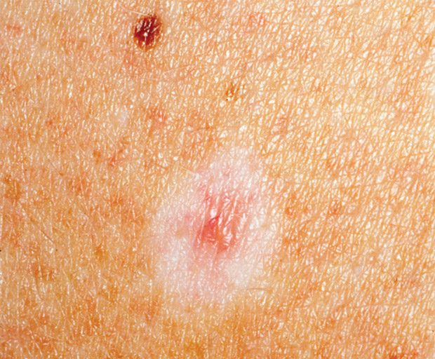
On physical examination, vital signs are normal. Skin findings are shown.
The remainder of the examination is unremarkable.
Which of the following is the most likely diagnosis?
A. Compound melanocytic nevus
B. Dysplastic nevus
C. Halo nevus
D. Malignant melanoma
Answer and critique
The correct answer is C. Halo nevus. This item is Question 50 in MKSAP 18's Dermatology section.
This patient has a halo nevus. Halo nevi are benign melanocytic nevi that are in the process of being attacked and eliminated by the immune system. They are characterized by a pigmented macule or papule with a surrounding “halo” of hypopigmented or depigmented skin. The halo gradually enlarges, and the mole shrinks until all that is left is a depigmented area on the skin that resembles a small area of vitiligo. These lesions are particularly common on the back in children, teenagers, and young adults.
Melanocytic nevi are benign neoplasms comprising melanocytes, commonly referred to as “moles.” They vary in clinical appearance depending on the location of melanocytes within the skin. Junctional nevi are flat and often dark brown in color. Compound nevi are raised (papules) and may be irregularly pigmented. Dermal nevi are soft and flesh-colored and may resemble skin tags. The terms “junctional,” “compound,” and “dermal” refer to the location of the melanocytic nests: junctional nevi have nests located at the dermal-epidermal junction, compound nevi have nests at the dermal-epidermal junction and in the dermis, and dermal nevi have nests in the dermis. Nevi undergo a benign maturation process from junctional to compound to dermal over a period of many years.
Dysplastic nevi can display one or more of the identifying characteristics of melanoma, making an accurate clinical diagnosis difficult; however, a biopsy of the lesion can typically exclude melanoma. They are often asymmetric, irregularly bordered, and more than one shade of brown, and they may be quite large in diameter. Many dysplastic nevi have a dark brown center and a surrounding ring of light brown, a pattern that is often referred to as a “fried egg” appearance. Patients with multiple dysplastic nevi are at risk for developing melanoma and should be monitored closely. Melanoma can arise within any melanocytic nevi including dysplastic nevi; therefore, biopsy should be limited to clinically worrisome or changing lesions.
Malignant melanoma presents as macules, papules, or plaques typically larger than 6 mm in diameter and shows asymmetry and irregular boarders, as well as color variation within the lesion. The surrounding skin can show erythema or hypopigmentation.
Key Point
- Halo nevi are benign pigmented macules or papules with a surrounding “halo” of hypopigmented or depigmented skin, most frequently presenting on the back of teenagers and young adults.